

Blepharitis
It is chronic inflammatory condition of eyelid margin. It occurs in two forms squamous and ulcerative type.
Squamous variety is more common and is observed in children especially those living in poor hygienic conditions.
It is many times associated with some form of refractive error similarly constitutional factors such as dandruff
over the scalp, excess of carbohydrate intake; digestive disturbances, allergic rhinitis & eczematous condition of
skin were also reported.
Patients usually represents with a chronic symptoms of appearance of white scales over the base of eyelashes in the
morning associated with itching and redness of eyes. There is a temporarily loss of eyelashes and lid margins are swollen
and has a reddish appearance.
Use of local treatment in the form of ointment helps to reduce the complaints, however its effect is temporarily as
well as some patients subsequently may develop allergic reaction to this local medication.
Use of alternative medicines like homoeopathy was found effective to give relieve in acute complaints of blepharitis.
Similarly constitutional homoeopathic treatment after detail history taking has helped in chronic cases of blepharitis.
Homoeopathic medicines being sweet and easy to administer children do take it regularly. Medicines are given in small dose
so it does not produce any untoward effects even if given for a long duration.
Case Reports :
A Case of Acute Blepharoconjunctivitis treated with Homoeopathy –

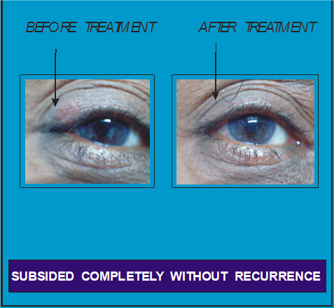
A case of Chronic Blepharitis treated with Homoeopathy
A female patient, aged 34 years came with complaints of itching of eyelid with whitish scales at the base of eye lashes for last 6 to 7 years.
It started in left eyelid and later on right lid was also affected. Patient was diagnosed as a case of chronic Blepharitis.
Conventional medicine in the form of ointment was prescribed by eye specialist.
It gave complete relief for a year or two but the same complaints started again.
So patient used the same ointment however it has not acted.
So patient started homoeopathic treatment that gave her relief in initial period but complaints started again.
So patient came to our institute and after detailed history taking homoeopathic treatment was given without using any local medication.
Patient’s took medicines regularly for 3 months and there was a gradual reduction of itching of lid and scales over eyelashes.
There was a complete relief of her complaints and no recurrence was noticed thereafter.

Chalazion
There is a development of a painless swelling in the eyelid. It is a swelling of meibomian gland following an obstruction of its duct,
accompanying by a chronic inflammation in the surrounding tarsus. [1]
The obstruction of the duct of gland is either due to proliferation
of its epithelium or by impaction of its secretion.
It occurs due to some endogenous infection, rosacea, poor lid hygiene, chronic blepharitis, seborrheic dermatitis, refractive error etc.
however many times no obvious cause can be found out.
Patients comes with either a small, painless, round swelling in the eyelid especially upper one or may presents with an acute pain in eyelid when
it gets infected. Painless lid swelling may cause a cosmetic disturbance to a patient while a large and centrally located chalazion gives pressure
over the cornea leading to blurred vision.
Although Chalazion is known to undergo spontaneous resolution, most of them becomes small after months but complete spontaneous resolution rarely occurs.
[2]
A Chalazion may sometimes break open through the upper eyelid and the palpebral conjunctiva and granulation tissue protrudes out causing irritation,
watering and even discharge if there is a secondary infection. [3]
Treatment in modern literature includes use of warm compresses and lid hygiene as a conservative treatment for a small painless Chalazion that may result in its
resolution within 1 month. Otherwise Intra-lesional injections of long acting steroids may help in resolution of small chalazion. If it doesn’t resolve or in case
of large Chalazion a surgical incision and curettage is the treatment of choice. Recurring Chalazion is treated by a subconjunctival total excision. In an acute
stage of inflammationit is treated by meticulous eyelid hygiene together with local and systemic antibiotics.
There are some limitations of conventional treatment such as chances of recurrence of Chalazion after incision and curettage surgery,
if curetting is not done properly. While with the use of intra-lesional injection of corticosteroid there are chances of ocular complication
such as inadvertent corneal penetration, traumatic cataract and serious complications of corticosteroid injection. If there are multiple and
small chalazion they are difficulty to remove surgically or with an intralesional corticosteroid injection. [4] Some patients may experience
sensitivity or a resistance to a systemic use of antibiotics or anti-inflammatory drugs.
So by taking into account above limitations of conventional treatment, a systemic use of Alternative medicines were used
and they were found effective in reducing acute painful chalazion as well as they helped to resolve completely long standing
painless chalazion and surgery can be prevented.
References :
1. Lyle TK, Cross AG. May and Worth’s Manual of Diseases of the Eye, 12th ed., London: Bailliere, Tindall and Cox; 1959: p. 69,70.
2. Sihota R, Tandon R. editors. Parsons' Diseases of the Eye. 20th ed. New Delhi: Elsevier; 1984: p. 427.
3. Dhanda RP, Kalevar V. A Text book of Clinical Ophthalmology. India: Vikas Publishing House Pvt. Ltd.; 1987: p. 127-28.
4. Hosal BM, Zilelioglu G. Ocular complication of intralesional corticosteroid injection of a chalazion. Eur J Ophthalmol. 2003;13: 798-99.
Refer to pdf article :
Clinical Study.pdf
Corneal Abscess
Cornea is an anterior transparent portion of outer wall of the eyeball. Corneal stromal abscess means a circumscribed collection of pus within the layers of the cornea.[1]
It is usually endogenous in origin and commonly found near the limbal blood vessels close to corneal margin. It is a form of marginal keratitis that occurs due endogenous
infections such as tuberculosis, syphilis, measles, or immunologically‑mediated diseases that includes phlyctenular keratitis, acne rosacea, meibomitis, and blepharitis.[2]
Corneal abscess usually extends to the outer surface to discharge the pus, and an ulcer is formed or it
may extend in both directions at the same time resulting in perforation of the cornea.[1]
Treatment in modern medicine is use of local and systemic antibiotics. However some patients may have an allergic reaction or a drug resistance to antibiotics.
Now a days almost every type of bacteria has become stronger and less responsive to antibiotic treatment when it is really needed.[3]
Homoeopathy has been treating infectious diseases successfully without giving any recurrence since pre-antibiotics era. In homoeopathy,
different medicines were mentioned for corneal abscess, but there was a lack of published case study or a research work
with documentary evidence about corneal abscess being treated with homoeopathy.
Systemic use of Homoeopathic medicines was found effective in such cases and a complete resolution of abscess
in 3 patients was observed without giving any recurrence during long term follow-up in these cases.
References :
1. Norton AB. Ophthalmic Diseases and Therapeutics. 3rd ed. New Delhi: B. Jain Publishers (P) Ltd.; 1987. p. 261‑2, 627.
2. Sihota R, Tandon R. editors. Parsons’ Diseases of the Eye. 20th ed. New Delhi: Elsevier; 1984. p. 186‑97.
3. Missouri Department of Health and Senior Services. FAQ’s. Available from: http://www.health
Refer to pdf article :
Case Report.pdf
Dacryocystitis
Lacrymal system consists of lachrymal gland which secretes tears while lachrymal drainage passage near nasal side of the eye drains the tears out of the eye.
Diseases of drainage passage are more common than of lachrymal gland. Drainage passage consists of lachrymal punctum, canaliculi, lachrymal sac and nasolacrimal
duct that open in back portion of nasal cavity as shown in figure. Blockage anywhere in this passage leads to overflow of tears called as Epiphora.
Epiphora in babies occurs due to congenital non-canalisation of nasolacrimal duct and natural canalization occurs between 3 months upto 1 year.
In adults epiphora is more common in females as this passage is anatomically small in diameter as well as hormonal changes during menstrual cycle causes swelling
in this passage leading to its obstruction. Similarly obstruction in passage can occur due to inflammation or any
growth affecting nose where nasolacrymal duct opens. Due to obstruction if tear drainage is affected then it remains stagnated
in lachrymal sac and gets secondary infected by bacteria causing inflammation of lachrymal
sac called as Dacryocystitis. This infection leads to mucopurulent discharge from the eye. If this infection remains for a longer time the drainage passage gets more obstructed.
In modern medicine use of local and systemic antibiotic are prescribed for dacryocystitis. A local massage to lachrymal sac is also advised to empty its infected material before putting eye drops.
Surgery is advised, if this condition recurs and DCR surgery is recommended in younger age group where sac is connected to nasal cavity while DCT surgery is done is done is old people where lachrymal sac is removed.
Some patients are sensitive to antibiotics and surgical procedure has its own complications. So alternatives medicines were used in this condition and they were found effective in controlling even
acute infection in lachrymal sac (dacryocystitis) without need to use antibiotic and anti-inflammatory drugs. Similarly they were found helpful to remove obstruction in drainage passage and prevent recurrence of infection in chronic cases.
Success Stories :
A case of Dacryocystitis in a baby
A mother brought her baby of 1 month age with a complaint of a swelling and redness between right eye and nose with a yellowish discharge from same eye for last 2 weeks. It was diagnosed as a case of acute dacryocystitis by eye specialist and local antibiotic eye drops were given; however there was no relief. So they came to the institute’s OPD for second opinion and to take alternative treatment.
On eye examination, there was a swelling between right eye and nose with a swollen eyelid and there was a redness over the swelling. Swelling over the lachrymal sac was hard, painful on touch and there was a greenish yellow discharge after pressure over the sac.
Homeopathic medicine was given every 2 hours for 2 days. Mother was instructed to prepare medicine daily by dissolving four medicated globules of medicine in one-fourth cup of lukewarm water. During every dose, she was advised to dip a teaspoon in medicated water and to put it on the tongue of a baby.
After 2 days, there was reduction of swelling, redness of skin over the sac and the discharge from the eye was reduced so the same medicine was continued with the same dose. After 3 days, swelling was further reduced and there was no redness and there was a reduction of hardness and pain on pressure. There was a reduction of discharge and its colour has changed from greenish yellow to white which suggest that there was a reduction of infection. Other Homeopathic other medicine was prescribed for the next 5 days. After 10 days follow-up, there was a further reduction of swelling and white discharge; so, the medicine was continued with the same dose. After 1 week, swelling and discharge subsided significantly but not stopped; so, the medicine was continued. After 10 days, there was a complete resolution of swelling and no discharge after pressure over the sac; so, the medicine was discontinued.
This case has shown effectiveness of homeopathy in acute dacryocystitis even in a baby of just one month age.
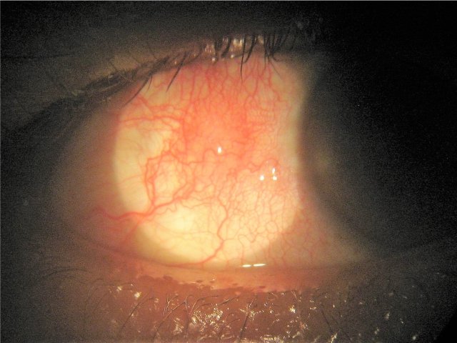
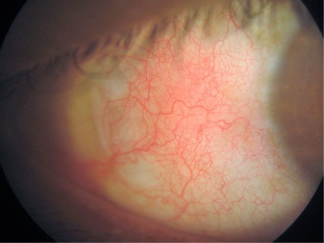
A case of acute dacryocystitis in a female aged 49 years :
A female aged 49 years reported on 26 March 2016, with a complaint of watering from right eyes for last 3 months. She also had a pain and swelling between the nose
and eye for last 2 days as shown in figure no.1. On examination swelling was painful and hard in consistency.
Patient was advised to apply cold application on the affected part and homoeopathic medicine was given by taking into account patients signs and symptoms.
After 4 days of treatment there was a reduction of pain, lid swelling, redness but hardness over swelling has not yet reduced please see figure 2. So other homoeopathic medicine was prescribed and after 3 days of follow-up there was a further reduction of swelling and pain as can be noticed from figure 3. After 4 days of continuation of medication, there was a complete resolution of swelling, pain and its hardness as can be observed from figure 4. Thus homoeopathy can help to reduced acute in inflammation without need to use allopathic medicines such as antibiotics and anti-inflammatory agents.


Myopia in Childhood
It is a common refractive error where an individual affected has dimness of vision for distant objects mainly due to increase in axial length of eyeball.
There are two varieties of axial myopia one is Simple (childhood) and other Pathological myopia. Out of these Childhood myopia is a common variety which
commences between 5 to 13 years of age, progresses during the period of body growth and after 25 years no major progression occurs.
Heredity does play an important role in etiology of myopia, however worldwide increased in its prevalence and progression during last few decades was attributed
to environmental factors especially excessive accommodation during near work associated with competitive education and urbanization. So controlling myopia is a burning
issue world over and lot of research is going on to control myopia.
Different measures used were vision therapy techniques, use of optical devices and local use of modern medicines. However these were not yet approved as a
gold standard either due to lack of their efficacy or safety or ethical issues associated with its long term use or rebound effect after discontinuation of treatment.
Whereas laser refractive surgery only corrects the existing myopia, so it is not recommended till myopia gets stabilise.
Clinical studies done so far in our institute has shown that use of alternative medicines has a controlling effect on childhood myopia,
similarly its effect continues even after stopping the treatment. Regression of myopia was also observed in few cases. However patient should take the medicines
regularly for the duration of one to two years, so that progression of myopia can be controlled and its stabilisation can be achieved.
Refer to pdf article :

Phlyctenular Conjunctivitis
It is an allergic reaction to endogenous bacterial proteins which in most cases are tuberculous but in other cases may be derived from mild,
long-standing infection such as those of tonsils and adenoids. Nowadays, its incidence has reduced may be due to improvement in diet and hygiene.
In this condition there is a formation of raised whitish yellow nodule on bulbar conjunctiva near or at the corneo-scleral junction (limbus) with a localised conjunctival congestion.
In modern medicine, systemic and local anti-inflammatory and antibiotics are used; however, there are chances of recurrence.
In homoeopathic literature different medicines were mentioned for this conditions deepening on the causation of Phlycten.
It was found that if Homoeopathic medicines were used according to aetiology of this condition, it gives quick results without need of any local medication and no recurrence was observed.
Refer to a documented case below
Case Report :
A Female aged 26 years reported on 17th November 2003 with complaints of redness and a raised spot in her left eye since 3 days and there was also yellowish bland discharge from the same eye for almost 3 weeks. On eye examination, there was a whitish yellow nodule in left eye on temporal side of conjunctiva near the limbus with a conjunctival congestion. Homeopathic medicine was prescribed four times a day for 4 days.
During treatment period, there was a gradual reduction of conjunctival congestion and discharge.
During eye examination, no visible nodule and congestion was noticed so medicine was discontinued. There was no recurrence of this condition after stopping the medicine.
This gives documentary proof about the effect of systemic homoeopathic treatment in phlyctenular conjunctivitis when a properly selected medicine is given.


Post Capsular Opacification
Posterior Capsular Opacification (PCO) is also known as 'after cataract’, which develops over the clear posterior capsule of a lens (a part of natural lens) a few months to a few years after an uneventful extracapsular cataract surgery.
It occurs as results of the growth and abnormal proliferation of lens epithelial cells on the posterior capsule. PCO remains the most common complication of cataract surgery, especially in young adults and children. It leads to gradual dimness of vision, glaring of light and eye strain while looking at near or distant objects.
It can be treated with either surgical intervention, such as posterior capsule scraping, or with a nonsurgical YAG laser capsulotomy. However, the latter is a costly procedure and has some clinical complications that include IOL optic damage, elevation of intraocular pressure, cystoid macular oedema and retinal detachment, as well as posterior capsulotomy can increase the risk of posterior segment complications especially in high myopes.
(Pandey SK, Apple DJ,Werner L, Maloof AJ, Milverton EJ. Posterior capsule opacification: a review of the aetiopathogenesis, experimental and clinical studies and factors for prevention. Indian J Ophthalmol 2004;52(2):99–112.)
Local and systemic uses of Homeopathic medicines were found helpful to promote the absorption of lens fragments after cataract operation. However patient has to take medicines for the duration of 4 to 6 months to get the results. So homeopathy can be used as a good option to treat such condition.

Pterygium
In this condition there is a formation of a fibro-vascular growth of conjunctiva (membrane that covers white portion of eye) over the cornea (central transparent portion) This growth occurs on the portion of eye which is exposed to atmosphere and it is common in individuals who are more involved in outdoor activity in dry, sandy and sunny atmosphere. So it is common in tropical areas.
Patient complaints of repeated redness, foreign body sensation, sticky discharge from eyes after going outside especially in summer season. Pterygium may cause a cosmetic disturbance especially if a patient is of younger age group. If this tissue growth continues to progress then it may involve central portion of cornea causing its distortion affecting vision drastically.
Treatment in early stages is conservative by use of sunglasses and lubricating eye drops. Treatment in modern medicine is concern, surgery is usually a treatment of choice for this condition, however post-operative recurrence with more rapid progression is a risk factor.
A retrospective clinical study was done in our institute in 27 patients and homoeopathic medicines were used by taking into account their eye complaints, eye examination findings and their general complaints. Medicines were given for the duration of 1 to 2 years depending on the response to treatment. Medicines were found effective in giving relief of symptoms in majority of the patients. They were also found helpful to reduce the thickening of pterygium, whereas regression of its head was observed in few cases. It has been observed that to achieve satisfactory results patient has to take medicines regularly as this condition is an end result of long term exposure of eyes to sunlight, dry and dusty atmosphere. No untoward effects of medicines were noticed during this study even though they were used for a long duration of 2 years.
Refer to pdf article :

Spring Catarrh
Spring Catarrh or Vernal Keratoconjunctivitis (VKC) is an inflammatory condition of conjunctival tissue of eye caused by allergy to exogenous allergens such as dust, heat; pollens etc. It occurs commonly in children & young adults. It leads to redness, itching, foreign body sensation in the eye and glaring of light. These complaints are common in summer season, however in tropical countries like India in some patients symptoms continue throughout the year. Majority of patients gives past history of atopy such as asthma, rhinitis and skin affections.
In modern medicine, use of local medicines especially steroids gives fast relief of complaints however their effect remains temporary and if used inadvertently on long term basis can leads to blinding complications such as corneal ulcer, cataract and glaucoma. Non-steroidal anti-inflammatory agents and mast cell stabilisers are also used in chronic cases. Although topical anti-allergic and anti-inflammatory eye drops are the mainstays of treatment for VKC, a gold standard treatment has not yet been established for this disease.
So by taking into account its chronicity, endogenous allergic nature and atopic background, a systemic approach of treatment with alternative medicine like Homoeopathy was considered instead of local treatment and observations of clinical study were as follows :
In acute stage of the disease medicines were found effective in giving symptomatic relief without need of any local medicines. In chronic cases, medicines were found useful to prevent the recurrence as well as helped to improve the general immunity of these patients. Homoeopathic medicines are sweet, easy to administer, so children prefer to take these medicines regularly.
Refer to pdf article :

Vitreous opacities
Vitreous humour is a transparent, colourless mass having a gel like consistency which fills the posterior cavity of eyeball behind the lens and maintenance the shape of eyeball. Vitreous Opacities (VO) are suspended matter in vitreous which vary in shape and size either in the form of fine spots, threads or appear as a membrane. They may remain fixed or float as the eye moves. Their colour varies from a gray to jet black.
Muscae volitans is the term used for appearance of spots before the eyes if there are no appreciable structural changes in vitreous or other media. Their causation has not been satisfactorily explained, but may be due to some imperfect incorporation of particles of transparent vitreous gel. They are found frequently in association with various general conditions such as weakness in anaemia, menstrual disturbances, digestive derangement etc. They are more appreciated when a patient looks against a plane background such as a wall or towards the sky. If they are big enough may cause a disturbance in vision especially during near work.
Vitreous opacities can occur due to liquefaction of vitreous in elderly persons. They occur frequently in myopes especially with posterior staphyloma and choroidal changes. They can be due to inflammation of ciliary body, choroid or retina as well as from vitreous haemorrhage.
Taking into account aetiology of vitreous opacity if alternative medicines are given then these opacities can be resolved completely as it was observed in many patients.
However during the course of treatment if a patient experience a sudden increase in opacities or an appearance of flashes of light in front of vision then it requires urgent examination by eye specialist as there are chances of development of retinal abnormalities such as retinal detachment especially in high myopic individuals or a posterior vitreous detachment in old individuals.
Success Story:
A patient aged 61 years, came with a complaint of appearance of a brownish colour spot in front of vision in left eye for last 4 to 5 years. He also experiences a web or a membrane in front of right eye since last 2 weeks. He is a known case of hypertension which is well controlled for last 5 years. He had associated complaints of allergic common cold. On eye examination, there were no obvious abnormalities except that he has to use presbyopic glasses for near work. By taking into account his ocular and nasal complaints, homoeopathic medicines were given and within one month opacity in right eye disappeared while after 7 months of medication opacity in left eye resolved completely and there was no recurrence thereafter.

Episcleritis
This is a condition where there is inflammation of deep subconjunctival connective tissue caused by an allergic reaction to an endogenous toxin. The episclera is a thin layer of tissue that lies between the conjunctiva and the connective tissue layer that forms the white of the eye (sclera). There are two forms of this condition: nodular and simple. Nodular episcleritis is characterized by a discrete, elevated area of inflamed episcleral tissue. In simple variety, there is a vascular congestion in a localized area without any obvious nodule.
Most cases of episcleritis are idiopathic. Approximately 26-36% of patients have an associated systemic disorder such as rheumatoid arthritis, colitis, gout, bacterial infections etc. It is common in young adults and in females.
Patient complaints of redness, mild pain in one or both eyes without discharge affecting one sector of the eye. It usually lasts for several days or weeks but has a tendency to recur.
In modern medicine topical steroids and nonsteroidal anti-inflammatory drugs are used, however they gives only temporary relief and there are chances of recurrence. Different medicines are mentioned for this condition in alternative therapy. Probable cause of nodular episcleritis has to be sort out after detail history taking, general examination and laboratory investigations.
If proper alternative medicine is selected depending on cause in a patient then this condition gets resolve with systemic medication without need of any local treatment. Similarly no recurrence was observed after systemic medication.
Success Story:
A male patient aged 62 years came in institute OPD on 12 July 2018 with the complaints of redness swelling and occasional foreign body sensation on his right eye.


He has used local eye drops from a doctor but there was no relief so came for alternative treatment. He gave history of undergone a root canal dental
treatment one year back. Patient had no complaint about tooth as well as no general complaints. He is a
known case of Hypertension and diabetes which is well controlled. There were no other abnormalities
detected in the eye as well as on general examination. Patient was explained about endogenous allergic
nature of this condition and systemic Homoeopathic medicine was started by taking into account his
previous dental treatment. No local medication was given.
After 1 week of treatment, redness of eye was reduced along with reduction of swelling and foreign
body sensation in eye. No recurrence of eye complaint was noticed by patient thereafter.


Diabetic Retinopathy
It is one of the major causes of blindness in the world. Those diabetic patients taking Insulin or those having uncontrolled diabetes are prone for development of diabetic retinopathy. Smoking and uncontrolled blood pressure are other risk factors that adversely affects diabetic retinopathy.
In this condition there occurs occlusion of capillaries of retina due to increase in viscosity of blood and stickiness of platelets which subsequently leads to reduction in blood supply of retina. In order to supply oxygen to hypoxic retina, new blood vessels are formed. There occurs an increase in vascular permeability and the overall effect is development of haemorrhages and oedema on retina. If these changes involve the central portion of retina (macula) then vision is affected drastically.
In modern medicine laser treatment is usually given to seal new bleeding vessels, while injections in the eye are given to treat macular oedema, however this is not a curative treatment, because this treatment requires repetition as well as it is not cost effective.
Cunha-Vaz has stated that “A number of large clinical trials have validated treatment methods now considered standard. However, the disease continues to progress in approximately 50% of the eyes treated by photocoagulation. Other forms of therapy targeted at the earliest stages of retinal disease are needed.”
(Cunha-Vaz J. G. Diabetic Retinopathy: Surrogate Outcomes for Drug Development for Diabetic Retinopathy. Ophthalmologica 2000;214:377–380.)
Intravitreal anti-VEGF injections are used in patients with diabetic macular edema, neovascular complications of diabetic retinopathy and retinal vascular occlusions. However there are reports of decrease in retrobulbar blood flow parameters, retinal arteriolar vasoconstriction, and worsening of macular ischemia after intravitreal injection of anti-VEGF agents.
(K Ghasemi Falavarjani, QD Nguyen. Adverse events and complications associated with intravitreal injection of anti-VEGF agents: a review of literature. Eye (2013) 27, 787–794.) So an alternative treatment was considered necessary for this condition.
Clinical study done in our institute :
Total 25 patients taking Ayurvedic treatment who came for regular follow-up for
5 years were studied retrospectively and following results were observed :
In 10 cases of Background Diabetic retinopathy there was no progression.
In 13 cases had pre-proliferative DR and undergone laser treatment for 2 or 3
times but still having microanurism were stablised after Ayurvedic treatment
and no more laser treatment was suggested by vitreoretinal surgeon.
In 2 cases had a proliferative DR and undergone panphotocoagulation laser
treatment but still had haemorrages on retina. After starting Ayurvedic
treatment haemorrhages was absorbed with an improvement in vision.
Thus Alternative medicines help to reduce obstructions in blood vessels
and improve blood circulation of retina. This helps to avoid formation
of new blood vessels on retina and patients don’t require to take laser
treatment or injections. These medicines can be used for long duration
without producing any untoward effects.
Documented Case reports :
Case 1 :
A female aged 56 years of age was suffering from Diabetes.
She was initially taking tablets to control her diabetes but later on shifted to Insulin.
She was diagnosed to have a Diabetic Retinopathy in 1993 and so started Ayurvedic treatment for the same.
Her retinal changes have not progressed, in fact regression was observed and no laser treatment was required thereafter.

Case 2 :
A male patient aged 45 years came with dimness of vision in left eye in 1999. His central portion of retina was affected and after starting Ayurvedic medicines for 2 years his vision improved to normal and not required to do a laser treatment.

Case 3 :
A patient was diagnosed to have a pre-proliferative retinopathy in 1998 with bleeding on retina for which laser treatment was given. Later on an Ayurvedic treatment was started and retinal condition remained stable and no more laser treatment was required thereafter.

Case 4 :
A patient was diagnosed to have a proliferative DR in 1993 where there were haemorrhages on retina and a scar tissue due to absorbed old haemorrhage. Ayurvedic treatment was started and it helped to absorb haemorrhages on retina. Patient was kept for follow-up up to the year 2005 and retinal condition remained stable.


Central Serous Retinopathy
It is a disease of retina where there is a collection of fluid in the macular portion of retina. Even though exact mechanism of development of CSR is not understood however many associations have been found. CSR is common in males between 20 to 50 years of age and those taking steroid medication or living in excessive mental stress.
Patients come with complaints of blurred or distorted vision usually affecting one eye only. It usually resolves spontaneously within 3 months. (Liew G, Quin G, Gillies M, Fraser-Bell S. Central serous chorioretinopathy: a review of epidemiology and pathophysiology. Clin Experiment Ophthalmol. 2013;41(2):201-214.)
Recurrences of CSR have been documented in up to 50% of patients within one year. (Aggio FB, Roisman L, Melo GB, Lavinsky D, Cardillo JA, Farah ME. Clinical Factors Related To Visual Outcome In Central Serous Chorioretinopathy: Retina. 2010;30(7):1128-1134.)
After resolution of CSR visual acuity will be regained, however in some patients some visual abnormality may remain.
Patients who were on steroid treatment are advised to taper down its dose and to stop later, while those having a stress, a psychosocial counselling are required. Alternative medicines were given in such cases and these medicines were found helpful to absorb the leakage as well as to reduce stress. These medicines also help to recover the vision quickly and no visual abnormality remains which is caused by scarring in retinal layers. Similarly these medicines help to prevent its recurrence in chronic case of CSR.
A Case Report :
A young patient aged 36 years came on 19th September 2014 with the complaint of dimness of vision in left eye for last 2 weeks. Before coming to our institute he consulted ophthalmologist and was diagnosed to have a CSR after undergoing OCT test. Figure 1 A & B shows OCT report showing macular oedema.


His vision recorded on Snellen’s chart was 6 / 36 in left eye, while it was normal (6 / 6) in right eye.
Ayurvedic treatment was planned after taking into account his history and eye examination findings. After 1 month of medication patients vision was improved to normal that is 6 / 6 in left eye on Snellen’s chart. [Figure 2 A & B] Medicines were continued for further 3 months. He was kept under follow-up for 1 year, however no recurrence was observed thereafter.


ARMD
ARMD is a common cause of irreversible blindness in patients above 50 years of age. There are two varieties of this disease dry and wet ARMD. Dry variety is common and causes a gradual dimness of vision due to degeneration of retinal pigment epithelial cells leading to pigmentation and eventually scar formation. Whereas in wet variety, there occurs a rapid loss of vision occur due to bleeding in macular area of retina from abnormal blood vessels growing from choroid.
Patient complaints of dimness of vision especially during near work and require more bright light. They also experience distortion in vision and straight line appears wavy as well as appearance of fogginess in central vision. Some patient may complaint of disturbances in colour vision.
Exact aetiology of ARMD is unknown and it is a disease of multifactorial in origin. Age is the greatest risk factor while other predisposing factors include heredity, smoking, obesity, exposure to UV radiation, diseases such as Hypertension, Diabetes and nutritional deficiencies.
Treatment in modern medicine includes use of Antioxidants however its usefulness is not proved. You can’t continue this treatment for life time due to its adversed effect.
In wet variety laser photocoagulation is used to seal new vessels but the results of large Randomised studies have shown that new vessel growth may occur at an increase rate after this treatment.
Anti- VGEF (vascular endothelial growth factor) treatment in the form of intra vitreal injections are used to control neovascularization, however they are not cost effective and its effect remains only for 1 to 2 months so has to be repeated. Despite encouraging results in halting the disease and improving the vision, repeated and long-term injections that are commonly needed may increase the chance of ocular and systemic complications. Ocular side effects include infection in the eye (Endophthalmitis), raise intraocular pressure (Glaucoma), ocular haemorrhages, retinal detachment etc.
(K Ghasemi Falavarjani and QD Nguye. Adverse events and complications associated
with intravitreal injection of anti-VEGF agents: a review of literature. Eye (2013) 27, 787–794.)
One large scale study has found out that repeated dosing of Intravitreal injection
of ranibizumab (Lucentis) can leads to retinal atrophy in 18 % cases who had no
atrophic changes during their enrollment in the study.
(Grunwald JE, Daniel E, Huang J, et al. Risk of geographic atrophy in the Comparison of Age-related Macular Degeneration Treatments Trials (CATT). Ophthalmology. 2013 Sep 30.)
Thus there is no satisfactory and safe treatment for ARMD in modern medicine. So alternative medicines like Ayurved and Homoeopathy offers a safe and effective approach of treatment for this condition.
Published Clinical Study Summary :
Total 25 cases of ARMD who took treatment in our institute were studied retrospectively.
Following observations were noted :
Good improvement was observed within a short period of treatment in dry variety of
ARMD which is confirmed by testing vision and on retinal examination.
In wet variety, haemorrhage and odema gets absorb after treatment with an
improvement in vision up to certain extent. Recurrence of haemorrhage gets arrested.
Patients should take treatment regularly to maintain the improved vision.
If macula is severely damaged with a scar formation then improvement is very limited.
Clinical Study :
Clinical StudyDocumented Case Report
A male patient came with a complaint of dimness of vision in both eyes and was diagnosed as a case of Dry ARMD in both eyes.
His vision was 6 / 60 in both eyes Fundus shows signs of Dry ARMD with geographical atrophic changes in right eye.
Ayurvedic treatment was started and after one year of treatment vision in left eye was improved to 6 / 9,
while in right eye no change was noticed probably due to atrophic changes.
 Check eye reports from eye specialist before and after treatment were shown in figure.
Similarly there was an improvement in Amsler’s grid test where wavy line are drawn by patient
(marked on grid) before treatment are shown in figure 6 and after treatment lines drawn by
patient on grid were more or less straight as can be observed from figure.
Check eye reports from eye specialist before and after treatment were shown in figure.
Similarly there was an improvement in Amsler’s grid test where wavy line are drawn by patient
(marked on grid) before treatment are shown in figure 6 and after treatment lines drawn by
patient on grid were more or less straight as can be observed from figure.


Ayurvedic treatment was planned after taking into account his history and eye examination findings. After 1 month of medication patients vision was improved to normal that is 6 / 6 in left eye on Snellen’s chart. Medicines were continued for further 3 months. He was kept under follow-up for 1 year, however no recurrence was observed thereafter.

Retinitis Pigmentosa
It is a degenerative condition of the retina causing of blindness in young adults. It is a caused by hereditary or it may occur in patients whose parents have married in close relation. Patients affected suffer from dim vision at night or in low light, loss of side (peripheral) vision, which may cause the person to bump into tables, furniture, or patients find difficulty in crossing the road.
Retinal examinations show appearance of black pigment along the vein and narrowing of blood vessels. In some patients macula is affected and optic disc becomes pale. Figure shows retinal appearance in RP affected individual.
Examination of visual fields and electrophysiological tests such as visual evoked potential (VEP) and electroretinogram (ERG) are helpful in diagnosis and assessing the progress of RP.
VEP is a noninvasive technique for assessing the function of visual system. In this method the eye is stimulated with a pattern stimulus and electrophysiological impulses were recorded using microprocessor controlled instrument known as Neuromatic 2000 C.
Despite of through studies in animals there is no rational or effective treatment for this condition in modern medicine.
Clinical study was done by giving Ayurvedic treatment and study summary is as follows :
Total 75 diagnosed patients suffering from primary pigmentary degeneration of retina (RP) were studied by taking detail history and eye examination includes testing vision, visual field and VEP.
VEP was done in normal individual of 25 males and 25 females to get normal data with Neuromatic 2000 C. [Figure 3]
Ayurvedic treatment was started and VEP test was carried out at base line and repeated after every 4 months. Similarly prothrombin time was checked at baseline.
Observations :
Deterioration in field of vision was arrested in all patients. There was slight improvement in vision noticed in 40 % cases. Improvement in night vision was observed in 2 cases.
Neuromatic 2000 C machine recording shows that there was an improvement in conduction time of optic tract as there was an improvement in signal to noise ratio. Similarly there was a marked improvement in formation of peaks in low and high intensity of light and a considerable improvement in latency values. [Figure 4] Refer figure 5, for latency values (conduction time measured in milliseconds for light to reach from eye to visual cortex in brain) in RP patients before and after treatment. Improvement was found rapid in children and young adults.
Prothrombin time was higher in RP patients as compared to normal individuals. Chromatographic study of plasma shows that in RP patients two binding proteins were absent as compared to normal individuals.


PUBLISHED RP STUDIES

Glaucoma
In this condition there is a gradual death or loss of retinal nerve cells and their fibres that makes optic nerve. There are mainly two varieties of glaucoma, open angle and close angle. Out of these open angle glaucoma is more common.
Open angle Glaucoma :
Damage to optic nerve is usually caused by rise in intraocular pressure (IOP) due to obstruction in drainage passage of a fluid (aqueous humour) whereas in some patients optic nerve gets damage even though the intraocular pressure is normal or low (low tension glaucoma). Abnormality in the circulation of the optic nerve or its deficient nourishment appears to be the reason for damage to the optic nerve. It is common after 40 years of age and many times associated with hypertension, diabetes and myopia. Severe psychological stress, drinking large amount of water as done in hydrotherapy can also leads to transient rise in intraocular ocular pressure.
In modern medicine initially eye drops are advised to control intraocular pressure (IOP) and if not found helpful to control pressure then surgery is advised. Systemic use of alternative medicines helps to lower ocular pressure by controlling excess formation of aqueous and reducing blockage in drainage passage of eye. This helps to improve peripheral vision to come extent which can be documented after examination of field of vision by autometed perimeter. In case of low tension glaucoma medicines helps to improve circulation and nourishment of optic nerve so that there occurs an improvement in vision and field of vision.
While taking alternative treatment patient should do a regular eye examination to check intraocular pressure and visual fields so as to know whether the medicine is working and to decide about its dosage.
Success story :
A male patient of 65 years age reported in our institute on 12 August 2016. He was a diagnosed case of Chronic simple glaucoma and undergone a combined Cataract with glaucoma surgery. However his ocular pressure was not under controlled, so he was on glaucoma eye drops as well as tablets (Diamox) for last 2 years. He is a known case of hypertension well controlled with medication for last 10 years. He gave history of pain in neck and tendency for constipation
Taking into account patients ocular and general complaints, Homoeopathic medicines were started and to my surprise within 2 weeks of treatment intraocular pressure came down as well as there was a reduction in myopia caused due to raised IOP. His eye specialist advised him to stop Diamox treatment and checked his pressure after 1 week which was still within their normal limits. Perimetry recording was also done and showed an improvement in visual fields in right eye while it was stable in the left eye.
After 7 months perimetry was repeated and there was stabilisation in field of vision. Similarly IOP was checked regularly and was found within in normal limits. Patient is still under homoeopathic treatment with a stable IOP.

Cataract
It is a condition where natural crystalline lens of eye starts becoming cloudy or opaque interfering with transmission of the rays of light into the eye and thereby causing a partial or total blindness.
It is common after 70 years of age, however heredity, exposure to UV radiation, prolong use of steroid medication, endogenous septic focus, injury, metabolic disturbances such as dehydration, diabetes as well as skin diseases makes people prone to develop cataract at an early age.
Cataract can be hard, soft or posterior sub-capsular. If it is a hard cataract patient requires bright light while doing near work or no longer requires his previous reading glasses for doing near work. In case of soft cataract, patient will have scattering of street light during night driving or double vision. If it is a posterior sub-capsular central it is many times central in position so it affects the vision drastically in bright illumination so patient will experience glaring of light.
Treatment of cataract is concerned; if the cataract is congenital that means right from birth and affecting vision then surgical treatment becomes necessary. Whereas cataract that occur in later life then we have to find out its cause by details history taking and give treatment accordingly. Now a days cataract surgery is advised routinely in a day to day practice. Surgical intervention leads to lot of financial burden over the nation, so there is a need to have some alternative treatment to arrest or slow down the progression of cataract. It was estimated that if the onset of cataract is delayed for 10 years, it will reduce the need of surgical intervention to half.
(Suryanarayana P, Saraswat M, Mrudula T, Krishna TP, Krishnaswamy K, Reddy GB. Curcumin and turmeric delay streptozotocin-induced diabetic cataract in rats. Invest Ophthalmol Vis Sci. 2005;46:2092–99)
However if cataract is in advanced stage, then it is not advisable to treat it medically but surgical intervention becomes necessary otherwise permanent visual loss can occur due to rise in intraocular pressure. (Glaucoma)
Local and systemic uses of Ayurved and Homoeopathic medicines if given in early stage of cataract are useful either in stabilization or to delay the progression of cataract. While taking alternative treatment patient should inform all his complaints to the doctor apart from their eye complaints so that it will help to know the cause of cataract and help in proper selection of medicine. While undergoing medical treatment for cataract patient is advised to check his eyes regularly so as to exclude any eye disease that arises with the old age.
From research work it was observed that intake of raw garlic in diet helps to delay the progression of cataract. Excessive consumption of sugar and salt can leads to development or rapid progression of cataract, so it is advisable to keep its consumption to minimum in your diet.